Fundus Diseases
2022-11-24 17:20:22
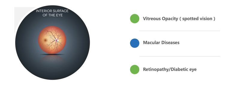
Vitreous Opacity(spotted vision) Macular Diseases Retinopathy/Diabetic eye
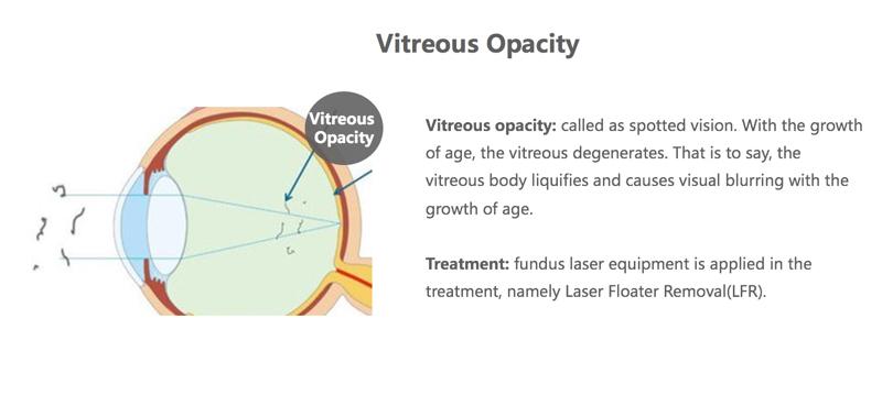
Vitreous Opacity Vitreous opacity: called as spotted vision. With the growth of age, the vitreous degenerates. That is to say, the vitreous body liquifies and causes visual blurring with the growth of age.
Treatment: fundus laser equipment is applied in the treatment, namely Laser Floater Removal(LFR).
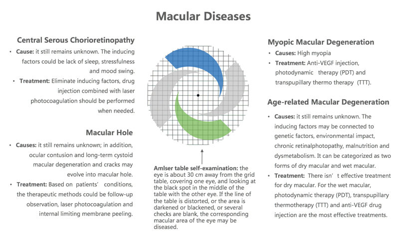
Macular Diseases Central Serous Chorioretinopathy Cause: it still remains unknown. The inducing factors could be lack of sleep, stressfulness and mood swing.
Treatment: Eliminate inducing factors, drug injection combined with laser photocoagulation should be performed when needed. Macular Hole Causes: it still remains unknown; in addition, ocular contusion and long-term cystoid macular degeneration and cracks may evolve into macular hole.
Treatment: Based on patients’ conditions, the therapeutic methods could be follow-up observation, laser photocoagulation and internal limiting membrane peeling.
Myopic Macular Degeneration Causes: High myopia
Treatment: Anti-VEGF injection, photodynamic therapy (PDT) and transpupillary thermo therapy (TTT). Age-related Macular Degeneration Causes: it still remains unknown. The inducing factors may be connected to genetic factors, environmental impact, chronic retinalphotopathy, malnutrition and dysmetabolism.It can be categorized as two forms of dry macular and wet macular.
Treatment: There isn’t effective treatment for dry macular.For the wet macular, photodynamic therapy (PDT), transpupillary thermotherapy (TTT) and anti-VEGF drug injection are the most effective treatments.
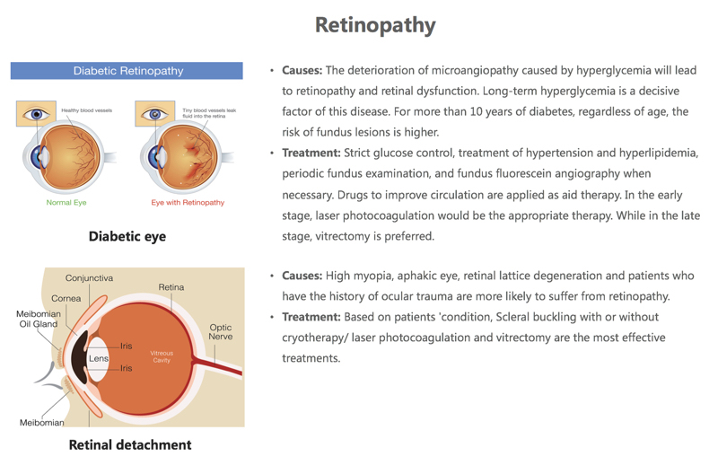
Retinopathy Causes: The deterioration of microangiopathy caused by hyperglycemia will lead to retinopathy and retinal dysfunction. Long-term hyperglycemia is a decisive factor of this disease. For more than 10 years of diabetes, regardless of age, the risk of fundus lesions is higher.
Treatment: Strict glucose control, treatment of hypertension and hyperlipidemia, periodic fundus examination, and fundus fluorescein angiography when necessary. Drugs to improve circulation are applied as aid therapy. In the early stage, laser photocoagulation would be the appropriate therapy. While in the late stage, vitrectomy is preferred. Causes: High myopia, aphakic eye, retinal lattice degeneration and patients who have the history of ocular trauma are more likely to suffer from retinopathy.
Treatment: Based on patients 'condition, Scleral buckling with or without cryotherapy/ laser photocoagulation and vitrectomy are the most effective treatments.
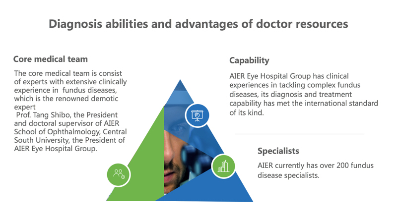
Core medical team The core medical team is consist of experts with extensive clinically experience in fundus diseases, which is the renowned demotic expert
Prof. Tang Shibo, the President and doctoral supervisor of AIER School of Ophthalmology, Central South University, the President of AIER Eye Hospital Group. Capability AIER Eye Hospital Group has clinical experiences in tackling complex fundus diseases, its diagnosis and treatment capability has met the international standard of its kind. Specialists AIER currently has over 200 fundus disease specialists.
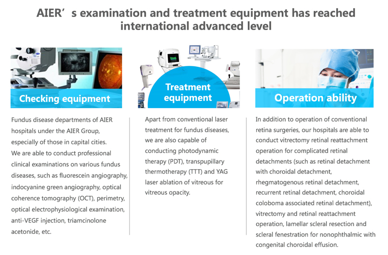
Fundus disease departments of AIER hospitals under the AIER Group, especially of those in capital cities.
We are able to conduct professional clinical examinations on various fundus diseases, such as fluorescein angiography, indocyanine green angiography, optical coherence tomography (OCT), perimetry, optical electrophysiological examination, anti-VEGF injection, triamcinolone acetonide, etc. Apart from conventional laser treatment for fundus diseases, we are also capable of conducting photodynamic therapy (PDT), transpupillary thermotherapy (TTT) and YAG laser ablation of vitreous for vitreous opacity. In addition to operation of conventional retina surgeries, our hospitals are able to conduct vitrectomy retinal reattachment operation for complicated retinal detachments (such as retinal detachment with choroidal detachment, rhegmatogenous retinal detachment, recurrent retinal detachment, choroidal coloboma associated retinal detachment), vitrectomy and retinal reattachment operation, lamellar scleral resection and scleral fenestration for nonophthalmic with congenital choroidal effusion.
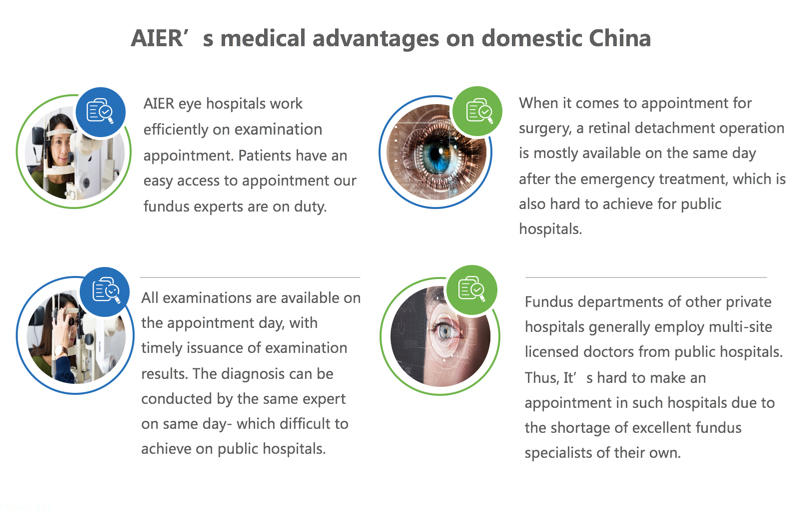
AIER eye hospitals work efficiently on examination appointment. Patients have an easy access to appointment our fundus experts are on duty.
When it comes to appointment for surgery, a retinal detachment operation is mostly available on the same day after the emergency treatment, which is also hard to achieve for public hospitals.
Fundus departments of other private hospitals generally employ multi-site licensed doctors from public hospitals. Thus, It’s hard to make an appointment in such hospitals due to the shortage of excellent fundus specialists of their own. All examinations are available on the appointment day, with timely issuance of examination results. The diagnosis can be conducted by the same expert on same day- which difficult to achieve on public hospitals.


